Instrument Hub
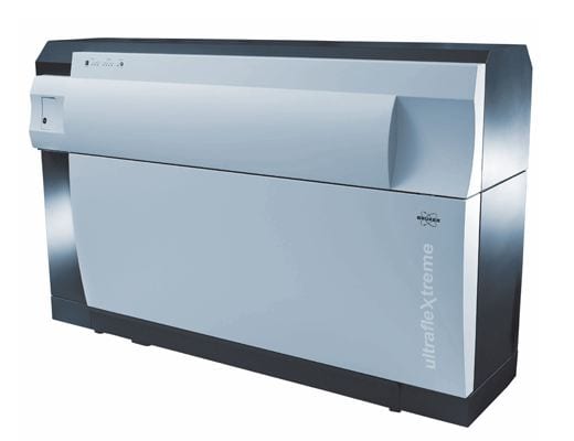
UltrafleXtreme
MALDI-TOF/TOF
Mass Spectrometer
The UltrafleXtreme enables you to analyse the mass of a wide variety of chemicals and biochemicals in either isolated spot preparations or in thin tissue sections with two dimensional spatial information.
Overview
The Biomedical Imaging Research Unit (BIRU) hosts a Bruker UltrafleXtreme MALDI-TOF/TOF mass spectrometer which can be used for standard MALDI analysis of purified biomolecules, chemical syntheses, or complex mixtures. Precursor ion analysis can be performed, in addition to MS/MS analysis for structural information.
The UltrafleXtreme also performs MALDI imaging mass spectrometry. In this experiment, a thin tissue section, evenly coated with MALDI matrix, is analysed by raster scanning the laser across the tissue in x and y. A mass spectrum is collected at each location, and the intensity of any detected m/z ratio plotted as a function of its location to generate two-dimensional MALDI images. In this way, a variety of biomolecules (proteins, peptides, lipids, metabolites, drugs, PTMs) can be spatially mapped with 10-250 μm resolution.
The UltrafleXtreme offers rapid mass analysis, with a wide range of molecules analysed (proteins, peptides, lipids, metabolites, drugs, polymers) over a wide mass range. It is especially suited for:
- MALDI imaging – either discovery or targetted analysis
- Manual or automated standard MALDI analysis
MALDI MS/MS analysisunfortunately our MS/MS capability is down – consider using the Bruker Solarix-XR for MALDI MS/MS- Posttranslational modification analysis
- Polymer analysis
Features
- 2 kHz Smartbeam II™ UV MALDI laser
- 10 micrometer laser beam size for MALDI imaging without pixel overlap
MS/MS analysis at 1 kHz laser repetition rate- Wide mass range resolving power up to 40,000 due to PAN™ Technology
- FlashDetector™ for 1 ppm mass accuracy
- Automated source cleaning with IR laser
Cost
Costs depend on nature of analysis – see the BIRU page for current information.
- Standard MS analysis – $23 per sample
MS/MS analysis – $10.50 per ion- Imaging analysis – $148.12/million laser shots
Eligibility
Post-graduate Students, Doctoral Candidates, Research/Academic Staff, Professional Staff, Collaborators (external to the University).
Access Requirements
Instrument is housed in the BIRU in a PC1 lab area, standard containment lab guidelines apply. Lab access requires health and safety training, and can be granted to users as applicable.
Training
The Instrument is not operated as a service. BIRU staff will train users on instrument operation, for standard MALDI analysis typically 1 x 2hr introductory session, and a further 1-2 hrs under supervision. For imaging analysis, in addition to introductory training, another 1 x 2hr session for imaging training and additional time for tissue preparation training is offered.
Booking
The UltrafleXtreme is booked through the BIRU online booking calendar once training has been received. Users need to first have hands on equipment training, lab orientation and containment training before they can book the UltrafleXtreme.
Software
Analysis of UltrafleXtreme data is supported by the following software:
- DataAnalysis
- FlexImaging
- SCiLs Lab 2D
Access to the above software is available on the MALDI Imaging or FT-ICR-MS Virtual Machines
Support
Email Gus Grey for all enquiries related to the UltrafleXtreme MALDI TOF/TOF.
Considerations
For standard MALDI analysis – presence of salts in buffers containing analytes should be avoided. For imaging analysis, fresh frozen tissue preferred, although some techniques for analysis of fixed/FFPE sections are possible. Sections should be 10-20 μm thick.
Data management and backup is generally the responsibility of the user. BIRU has an IT manager who can assist with data management issues.
Limitations
- Imaging spatial resolution is limited to 10 μm – and only for very abundant signals
- Imaging not suitable for low abundance protein analysis, membrane proteins/channels/transporters, or for analysis of intact proteins larger than ~50 kDa