MALDI Imaging Mass Spectrometry to validate novel transgenic mouse models lacking melanocortin peptide hormones
A/P Kathy Mountjoy and Dr Gus Grey, Department of Physiology, SMS, FMHSCollaborator: Dr Joel Elmquist, Centre for Hypothalamic Research, University of Texas Southwestern Medical Center, Dallas
Seeking: BSc (Hons), BBiomed Hons, Masters and PhD students with a passion for multidisciplinary biomedical research.
Introduction
________________________________________
Multiple melanocortin peptides in the pituitary and brain are some of the most important hormones involved in the regulation of stress, body weight, obesity, type-2 diabetes and neurological disorders. Despite these pivotal roles, very little is known about which of the specific roles for brain or pituitary derived melanocortin peptide hormones. We hypothesise that specific melanocortin peptides produced from brain and pituitary work together regulating diverse physiological responses. Specific functions for pituitary and brain melanocortin peptides are currently not understood. Here, we will utilise the specificity and sensitivity of imaging mass spectrometry to detect, map and quantify endogenous melanocortin peptide hormones in mouse pituitary and brain sections.
Objective
________________________________________
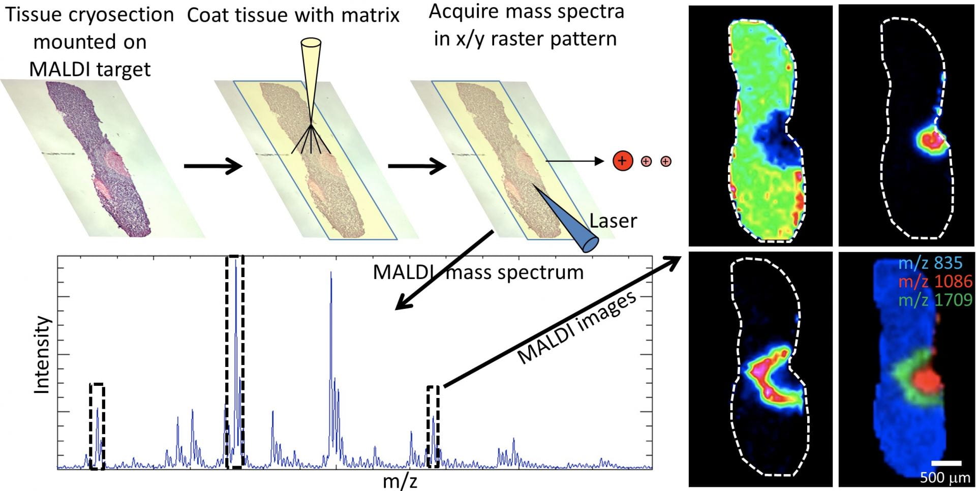
This project will apply state-of-the-art MALDI imaging mass spectrometry technology to identify, map, and quantitate POMC cleavage products through mouse pituitary and brain. MALDI imaging mass spectrometry is a proteomic technique that combines the specificity and sensitivity of mass spectrometry with spatial information to map tissue-wide distribution of multiple analytes simultaneously, directly from a single tissue section. A standard curve-based quantitative approach was used to generate proof-of-principle absolute quantitative MALDI images of POMC-derived peptides. Unique transgenic mouse models that lack specific melanocortin peptides either in the brain or in pituitary are being developed in Dallas. Frozen tissues will be sent to NZ and melanocortin peptide expression will be validated in this project using MALDI imaging mass spectrometry.
Significance
_______________________________________
This study will advance knowledge on how the natural melanocortin system regulates appetite and body weight. This knowledge could eventually lead to effective therapies for today’s obesity epidemic.
To apply, go to:
https://www.findathesis.auckland.ac.nz/research-entry/10467818
or contact: Dr Kathy Mountjoy (kmountjoy@auckland.ac.nz) or Dr Gus Grey (ac.grey@auckland.ac.nz)
________________________________________
Tissue cryosections are collected and prepared for MALDI mass spectrometry with 10-200μm spatial sampling. Lipids (A) and peptide hormones (B, C) were detected in specific lobes of the mouse pituitary.

